研究活動について
組織球症に伴う中枢神経変性症の診療ガイド2疾患概念
組織球症(histiocytosis: HC)は、単球系細胞が様々な臓器に集簇し、臓器傷害をきたす疾患の総称である。その中で、集簇する組織球にがん遺伝子変異を認める疾患群を組織球性腫瘍と呼び、小児に好発するランゲルハンス細胞組織球症(Langerhans cell histiocytosis: LCH)や黄色肉芽腫(xanthogranuloma: XG)、若年成人に多いロサイ・ドルフマン病(Rosai-Dorfman disease: RDD)や中高年に好発するエルドハイム・チェスター病(Erdheim-Chester disease: ECD)などが含まれる1)。(表1)これらは、腫瘍と炎症の両者の特性を持つため、炎症性骨髄腫瘍とも呼ばれる。
表1. 主な組織球性腫瘍
| 疾患名 | 好発年齢 | 病変/症状・特徴 |
|---|---|---|
| ランゲルハンス細胞組織球症 (Langerhans Cell Histiocytosis: LCH) |
主に乳幼児 (成人にも) | 溶骨、皮疹、リンパ節腫脹、肝脾腫、尿崩症など |
| 黄色肉芽腫 (XanthoGranuloma: XG) |
乳幼児 (成人にも) | 皮疹、眼・肺・肝臓・頭蓋内病変など |
| エルドハイム・チェスター病 (Erdheim-Chester Disease: ECD) |
中高年 | 両側脛の骨硬化、心臓・大血管・腎周囲の線維化病変など |
| ロサイ・ドルフマン・デトン病 (Rosai-Dorfman-Destombes Disease: RDD) |
主に若年成人 | 両側頸部のリンパ節腫脹、皮膚・骨・副鼻腔・眼窩病変など |
| 不確定樹状細胞組織球症 (Indeterminate Dendritic Cell Histiocytosis: IDCH) |
中高年 | 皮疹病変が主、他の血液腫瘍を合併することが多い |
| 悪性組織球性腫瘍 (Malignant Histiocytic Neoplasms: MHN) |
主に成人 | リンパ節腫脹、脾腫、皮膚・肺・肝臓病変など、死亡率高い |
| 混合性組織球症 (Mixed Histiocytosis: MH) |
主に成人 | LCH+ECDが最も多く、次いでECD+RDD |
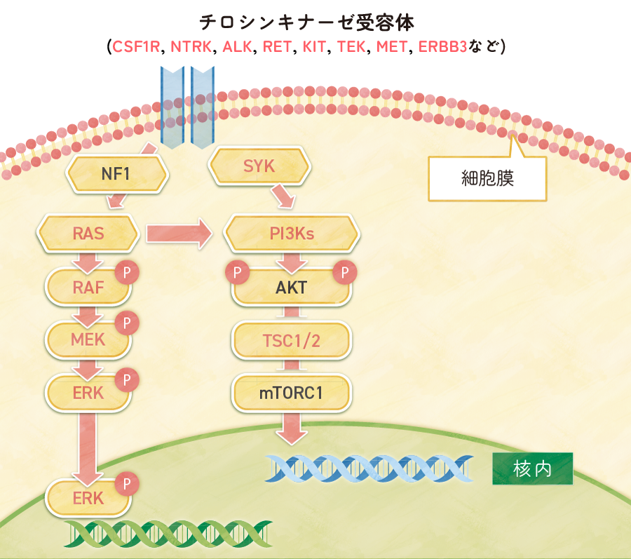
2010年にLCHの約半数の例で病変組織にBRAFV600E変異が検出されることが報告され2)、これを契機にMAP2K1などMAPK経路の遺伝子に活性化変異が次々に同定された。LCH患者の90%にMAPK経路に活性化遺伝子変異を認め3)、この変異はドライバー変異である4)。ECD患者にも半数近くにBRAFV600E変異を認め5)、NRASやKRAS等を含め90%にMAPK経路に活性化遺伝子変異が検出される4)。(図1)
図1. 組織球症腫瘍における遺伝子変異
LCHにおいて、LCH発症から数年以上経過し、腫瘍性病変が消失した時期に、小脳失調や高次脳機能障害が非可逆的に進行する中枢神経変性症(neurodegenerative disease: ND)が続発することが、1990年代前半から知られていた6)。ECDにおいても、診断時から徐々に進行する同様のNDの存在が、1990年代半ばから知られていた7)。最近、XGにおいても、同様のNDが報告されている8)。
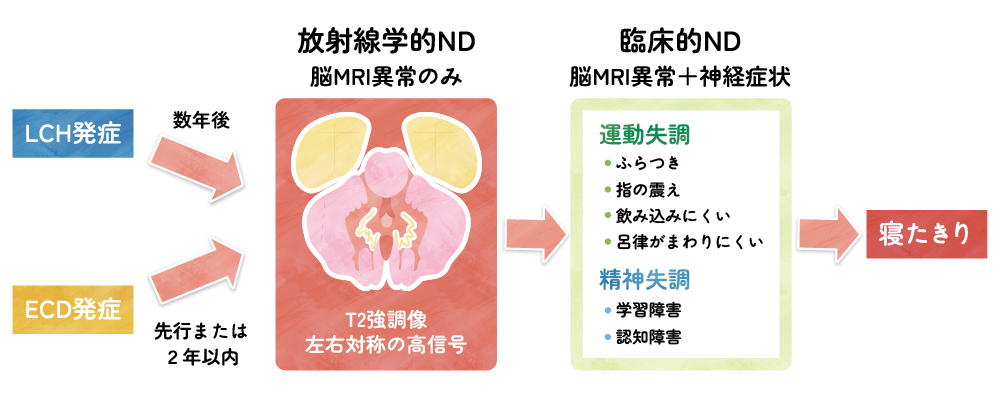
LCH診断時にLCH関連ND(LCH-ND)を認めることは極めてまれである。多くの場合、LCH診断後3年以上経過し、初期の腫瘍性病変が消失した時期に、脳MRIで小脳歯状核・基底核・橋にT2高信号の左右対称性の造影効果のない異常信号が出現・進行し9)、次第に進行して小脳や大脳の萎縮が現れる10,11)。MRI異常のある例の25%は、数年以内に小脳失調などの運動障害や、知能低下、学習障害、性格変化などの高次脳機能障害が出現する9,12)。(図2)
ECD関連ND(ECD-ND)においても、脳MRIでLCH-NDと同様の所見を認めるが、LCH-NDより早期に発現する。脳MRI異常や神経症状が出現する時期は、ECD診断に先行または同時が60%近く、ECD診断から2年以内が30%近くを占める。ECD-ND患者の80%に、神経症状出現時に骨病変や後腹膜線維症などのECDの活動性病変を認める13,14)。(図2)
図2. LCH関連中枢神経変性症の典型的な経過
JXG関連ND(JXG-ND)の報告は1報(2例)4)しかなく、1例はJXG診断時に脳MRI異常を認めその数年後に神経症状を発症した。もう1例は再発時に一過性に脳MRI異常と神経症状を認めた。
RDD関連ND(RDD-ND)の報告はリンパ節型に合併した1例のみであり、リンパ節腫大に先行して小脳症状が出現していた15)。
参考文献
- McClain KL, Bigenwald C, Collin M, et al. Histiocytic disorders. Nat Rev Dis Primers. 2021; 7: 73.
- Badalian-Very G, Vergilio JA, Degar BA, et al. Recurrent BRAF mutations in Langerhans cell histiocytosis. Blood 2010; 116: 1919-1923.
- Durham BH, Lopez Rodrigo E, Picarsic J, et al. Activating mutations in CSF1R and additional receptor tyrosine kinases in histiocytic neoplasms. Nat Med 2019; 25: 1839-1842.
- Berres ML, Lim KP, Peters T, et al. BRAF-V600E expression in precursor versus differentiated dendritic cells defines clinically distinct LCH risk groups. J Exp Med. 2014; 211: 669-683.
- Haroche J, Charlotte F, Arnaud L, et al. High prevalence of BRAF V600E mutations in Erdheim-Chester disease but not in other non-Langerhans cell histiocytoses. Blood. 2012; 120: 2700-2703.
- Grois N, Barkovich AJ, Rosenau W, et al. Central nervous system disease associated with Langerhans' cell histiocytosis. Am J Pediatr Hematol Oncol 1993; 15: 245-254.
- Fukazawa T, Tsukishima E, Sasaki H, et al. Erdheim-Chester disease and slowly progressive cerebellar dysfunction. J Neurol Neurosurg Psychiatry. 1995; 58: 238-240.
- Daifu T, Umeda K, Yokoyama A, et al. Juvenile xanthogranuloma manifesting with LCH-associated neurodegenerative disease-like radiological findings. Pediatr Blood Cancer. 2024;71:e31043.
- Wnorowski M, Prosch H, Prayer D, et al. Pattern and course of neurodegeneration in Langerhans cell histiocytosis. J Pediatr 2008; 153: 127-132.
- Martin-Duverneuil N, Idbaih A, Hoang-Xuan K, et al. MRI features of neurodegenerative Langerhans cell histiocytosis. Eur Radiol. 2006; 16: 2074-2082.
- Prosch H, Grois N, Wnorowski M, et al. Long-term MR imaging course of neurodegenerative Langerhans cell histiocytosis. AJNR Am J Neuroradiol 2007;28:1022-1028.
- Heritier S, Barkaoui MA, Miron J, et al. Incidence and risk factors for clinical neurodegenerative Langerhans cell histiocytosis: a longitudinal cohort study. Br J Haematol 2018; 183: 608-617.
- Chiapparini L, Cavalli G, Langella T, et al. Adult leukoencephalopathies with prominent infratentorial involvement can be caused by Erdheim-Chester disease. J Neurol. 2018; 265: 273-284.
- Riso V, Nicoletti TF, Rossi S, et al. Neurological Erdheim-Chester Disease Manifesting with Subacute or Progressive Cerebellar Ataxia: Novel Case Series and Review of the Literature. Brain Sci. 2022; 13: 26.
- Candeias da Silva C, Pedroso JL, Moraes FM, et al. Teaching NeuroImages: Rosai-Dorfman disease presenting with progressive early-onset cerebellar ataxia. Neurology. 2013; 81: e27-28.

本研究は、AMEDの課題番号JP23ek0109635の支援を受けています。